❝Never do anything by halves if you want to get away with it. Be outrageous. Go the whole hog. Make sure everything you do is so completely crazy it’s unbelievable.❞
-Roald Dahl, Matilda (1988)

A ‘med comms’ site for everyone!

✒︎ Informative

Presenting the latest news, discoveries and innovations in medicine in a blog-style format!

⚕ Global

Reaches the national and the international readers!

♛ Unique & Dynamic

There’s an article for everyone in all areas of medicine!

#scriveners

The Plague of Ashdod (1630) Nicholas Poussin

The artwork “The Plague of Ashdod” was created by the French painter Nicolas Poussin in 1630. It portrays the biblical narrative of a divine plague inflicted upon the people of Ashdod. 

This dramatic scene of divine punishment is described in the Old Testament. The Philistines are stricken with plague in their city of Ashdod because they have stolen the Ark of the Covenant from the Israelites and placed it in their pagan temple. You can see the decorated golden casket of the Ark between the pillars of the temple. People look around in horror at their dead and dying companions. One man leans over the corpses of his wife and child and covers his nose to avoid the stench. Rats scurry towards the bodies. The broken statue of their deity, Dagon, and the tumbled down stone column further convey the Philistines’ downfall.

In the artwork, Poussin vividly depicts the turmoil and suffering caused by the plague. The foreground is filled with the stricken inhabitants of Ashdod; their bodies are contorted in agony or limp in the stillness of death, illustrating the mercilessness of the affliction. The variety of postures and expressions captures the range of human suffering and chaos that accompanies such disaster. 

Amongst the afflicted, several figures stand out due to their dynamic gestures or central placement within the composition, drawing the viewer’s eye and emphasizing the emotional impact of the scene. In the background, classical architecture gives a sense of order and permanence that starkly contrasts with the disarray and despair of the figures. Poussin’s use of colour and light skilfully highlights the drama, with the dark and earthy tones of the suffering masses set against the lighter, more serene sky, which suggests divine presence or intervention.

Poussin’s use of color and light skillfully highlights the drama, with the dark and earthy tones of the suffering masses set against the lighter, more serene sky, which suggests divine presence or intervention. The overall effect is one of a carefully structured scene that conveys a narrative full of intensity and profound human drama, characteristic of the religious paintings of the period and the classical style Poussin is renowned for. Poussin began to paint The Plague of Ashdod while the bubonic plague was still raging throughout Italy though sparing Rome. He first called the painting The Miracle in the Temple of Dagon, but later it became known as The Plague of Ashdod.

The painting most importantly provides a view into how illness and diseases were feared at that time in the past and the fact that people had the knowledge that it was transmissible during that time period which was the 16th century.

𝐀𝐧𝐧𝐨𝐮𝐧𝐜𝐞𝐦𝐞𝐧𝐭: 𝐂𝐞𝐥𝐞𝐛𝐫𝐚𝐭𝐢𝐧𝐠 𝟐𝟎𝟎 𝐩𝐨𝐬𝐭𝐬 𝐦𝐢𝐥𝐞𝐬𝐭𝐨𝐧𝐞 𝐫𝐞𝐚𝐜𝐡! 𝐈 𝐜𝐚𝐧’𝐭 𝐭𝐡𝐚𝐧𝐤 𝐞𝐚𝐜𝐡 𝐨𝐧𝐞 𝐨𝐟 𝐲𝐨𝐮 𝐞𝐧𝐨𝐮𝐠𝐡! 𝐖𝐞’𝐫𝐞 𝐚𝐭 𝐚 𝟓𝐤 𝐬𝐭𝐫𝐞𝐚𝐤 𝐚𝐬 𝐰𝐞𝐥𝐥! ♥️🍾🍷#scriveners
𝘗𝘭𝘦𝘢𝘴𝘦 𝘤𝘩𝘦𝘤𝘬 𝘰𝘶𝘵 𝘰𝘶𝘳 𝘯𝘦𝘸𝘭𝘺 𝘶𝘱𝘥𝘢𝘵𝘦𝘥 ‘𝘌𝘹𝘵𝘳𝘢𝘴 𝘗𝘢𝘨𝘦’!╰(°▽°)╯
𝕸𝖊𝖗𝖗𝖞 𝕮𝖍𝖗𝖎𝖘𝖙𝖒𝖆𝖘!🎄🎅𝕸𝖆𝖞 𝖆𝖑𝖑 𝖞𝖔𝖚𝖗 𝕮𝖍𝖗𝖎𝖘𝖙𝖒𝖆𝖘 𝖜𝖎𝖘𝖍𝖊𝖘 𝖈𝖔𝖒𝖊 𝖙𝖗𝖚𝖊!

🥳𝐉𝐮𝐬𝐭 𝐢𝐧𝐬𝐭𝐚𝐥𝐥𝐞𝐝 𝐚 𝐧𝐞𝐰 𝐩𝐥𝐚𝐧 𝐚𝐧𝐝 𝐜𝐡𝐚𝐧𝐠𝐞𝐝 𝐭𝐡𝐞 𝐬𝐢𝐭𝐞 𝐚𝐝𝐝𝐫𝐞𝐬𝐬! 𝐖𝐞’𝐯𝐞 𝐮𝐩𝐠𝐫𝐚𝐝𝐞𝐝 𝐛𝐚𝐛𝐲! 🎉 scrionl.blog ♡
🚨𝐃𝐮𝐞 𝐭𝐨 𝐬𝐨𝐦𝐞 𝐮𝐧𝐟𝐨𝐫𝐞𝐬𝐞𝐞𝐧 𝐜𝐢𝐫𝐜𝐮𝐦𝐬𝐭𝐚𝐧𝐜𝐞 𝐈 𝐰𝐢𝐥𝐥 𝐛𝐞 𝐭𝐚𝐤𝐢𝐧𝐠 𝐚 𝐡𝐢𝐚𝐭𝐮𝐬 𝐟𝐨𝐫 𝐚 𝐩𝐞𝐫𝐢𝐨𝐝 𝐨𝐟 𝐨𝐧𝐞 𝐦𝐨𝐧𝐭𝐡!🚨
𝐖𝐞 𝐧𝐨𝐰 𝐡𝐚𝐯𝐞 𝐚𝐧 𝐈𝐧𝐬𝐭𝐚𝐠𝐫𝐚𝐦 𝐚𝐜𝐜𝐨𝐮𝐧𝐭!📱
𝐀 𝐧𝐞𝐰 𝐬𝐞𝐜𝐭𝐢𝐨𝐧 ‘𝐂𝐨𝐧𝐭𝐚𝐜𝐭’ 𝐡𝐚𝐬 𝐛𝐞𝐞𝐧 𝐚𝐝𝐝𝐞𝐝! 📞

𝐓𝐡𝐞 ‘𝐋𝐢𝐧𝐤𝐬 & 𝐁𝐨𝐨𝐤𝐬 & 𝐘𝐨𝐮𝐓𝐮𝐛𝐞 & 𝐏𝐨𝐝𝐜𝐚𝐬𝐭𝐬’ 𝐬𝐞𝐜𝐭𝐢𝐨𝐧 𝐢𝐬 𝐧𝐨𝐰 𝐚𝐯𝐚𝐢𝐥𝐚𝐛𝐥𝐞!💙
𝐍𝐞𝐰 𝐰𝐚𝐥𝐥𝐩𝐚𝐩𝐞𝐫𝐬 𝐡𝐚𝐯𝐞 𝐛𝐞𝐞𝐧 𝐚𝐝𝐝𝐞𝐝 𝐭𝐨 𝐭𝐡𝐞 ‘𝐄𝐱𝐭𝐫𝐚𝐬’ 𝐬𝐞𝐜𝐭𝐢𝐨𝐧. 𝐃𝐨 𝐜𝐡𝐞𝐜𝐤 𝐢𝐭 𝐨𝐮𝐭!⚡️
𝐀𝐧𝐧𝐨𝐮𝐧𝐜𝐞𝐦𝐞𝐧𝐭: 𝐌𝐨𝐫𝐞 𝐭𝐡𝐚𝐧 𝐚 𝟏𝟎𝟎 𝐭𝐡𝐚𝐧𝐤𝐬! 𝐖𝐞’𝐯𝐞 𝐫𝐞𝐚𝐜𝐡𝐞𝐝 𝟏𝟎𝟎 𝐩𝐨𝐬𝐭𝐬! 🍾 🍷
𝓒𝓮𝓵𝓮𝓫𝓻𝓪𝓽𝓲𝓷𝓰 𝓽𝓱𝓲𝓼 𝓶𝓮𝓭𝓲𝓬𝓪𝓵 𝔀𝓻𝓲𝓽𝓲𝓷𝓰 𝓫𝓵𝓸𝓰’𝓼 1-𝔂𝓮𝓪𝓻 𝓪𝓷𝓷𝓲𝓿𝓮𝓻𝓼𝓪𝓻𝔂!🍾🍷

Exploring the Meaning of Medical Anthropology, Conducting Research on Indigenous Shamans of Peru, and Notes on the Cholera Epidemic Part 3

Some treat their curing work as a divine calling; others seem more like pragmatic businessmen. Yet, there are striking similarities in the paths that led these exceptional individuals to the occupation of curandero, and there is an underlying curing philosophy that unites them. Our primary objective is ethnographic, to record in detail the symbolic system…

Exploring the Meaning of Medical Anthropology, Conducting Research on Indigenous Shamans of Peru, and Notes on the Cholera Epidemic Part 2

Evolutionary and Ecological Perspectives on a Cholera Epidemic Medical anthropology theory is a blend of social science, epidemiological, and biological perspectives on disease. The most effective way to show how these theoretical threads come together in medical anthropology is to introduce them separately. Theories operate at an abstract level. They reduce the complexity of a…

Exploring the Meaning of Medical Anthropology, Conducting Research on Indigenous Shamans of Peru, and Notes on the Cholera Epidemic Part 1

North coast of Peru, 1980: It’s three in the morning, the ocean breeze is wet and cold. It is not clear if my present nausea is attributable to the mescaline-bearing San Pedro (Trichocereus pachanoi) brew with which I began the evening or to the vile black-leaf tobacco sludge I have been asked to snort repeatedly.…

Improving Survival Rates Against ALS and FTD

Being a medical student and having ALS is a toxic concoction of health longevity. Yentli Soto Albrecht is a researcher on ALS at Penn Medicine alongside her battles with the neuro-degenerative disease. She was genetically tested for having the disease and lost her 66 year old father to the same disease in 2024. Her goal…

An Online 3D Atlas of Human Organs

The Human Organ Atlas is made possible by funding from: The European Synchrotron Radiation Facility (ESRF), The Chan Zuckerberg Initiative, The German Registry of COVID-19 Autopsies (DeRegCOVID), supported by the German Federal Ministry of Health, The UK Medical Research Council (MRC), The Royal Academy of Engineering, The Wellcome Trust. The Human Organ Atlas (HOA), an…

The Unprecedented and Undisputed Wave of AI Doctors

This is Doctor Kevin Adewale, he wants you to know that he’s not an all-bs cardiac surgeon, he’s probably from Kenya but he could be Sudanese or Jamaican whatever your eyes want you to believe in… hmm he’s also got a strong following, a pretty fancy YouTuber thing he’s got going on. He’s yet to…

  • On Developing a Treatment for River Blindness 

    by

    Nivea Vaz ,
    5–7 minutes

    ‘Thanks to K-MEDI’s expertise and RIGHT Foundation support, we are hopeful we can deliver urgently needed treatments to the millions of people affected by river blindness. We need to address the gap in current treatments, and developing effective, targeted test-and-treat approaches with macrofilaricidal drugs like oxfendazole will be critical in reaching the WHO’s goal of eliminating this terrible and debilitating disease.’

    ~Dr Luis Pizarro, Executive Director of DNDi


    K-MEDI hub and the Drugs for Neglected Disease initiative (DNDi) have collaborated together in efforts to develop a treatment for onchocerciasis, the second leading cause of blindness after trachoma. Onchocerca volvulus is a type of roundworm called a filarial worm. There are 19 million people infected by the roundworm and this includes the 1.15 million people who have lost their eyesight!


    DNDi will be the principal investigator and K-MEDI is the collaborative institution. This R&D initiative is supported by the Research Investment for Global Health Technology Foundation (RIGHT Foundation), with a total of KRW 3.2 billion (approx. EUR 2.2 million) over a period of 2.5 years. Established in 2003 by Médecins Sans Frontières (MSF), Institut Pasteur, and four leading medical research institutes in neglected disease-endemic countries, DNDi is a non-profit international research organization dedicated to developing new treatments for neglected diseases. The molecule oxfendazole is going to be a safe, an effective, an affordable and a globally accessible treatment for the filarial worm.


    The RIGHT Foundation is the first international public-private partnership funding agency in Korea, jointly established in 2018 by the Ministry of Health and Welfare, the Gates Foundation, and Korean life science companies. It supports R&D projects that help alleviate the disproportionate burden of infectious diseases on low- and middle-income countries (LMICs), thereby contributing to global health equity. To date, the foundation has supported 70 research projects with a total funding of 107.7 billion KRW.


    For context…


    Onchocerciasis is the second leading cause of infectious blindness worldwide (after trachoma).


    Onchocerciasis is most common in tropical and sub-Saharan regions of Africa. Small foci exist in Yemen and along the Venezuelan border with the Brazilian Amazon. Blindness due to onchocerciasis is fairly rare in the Americas; Colombia, Ecuador, Mexico, and Guatemala have been declared free of onchocerciasis by the World Health Organization (WHO). People who live or work near rapidly flowing streams or rivers are the most likely to be infected. In addition to residents, long-term travelers (eg, missionaries, aid workers, field researchers) are at risk.


    Pathophysiology of Onchocerciasis


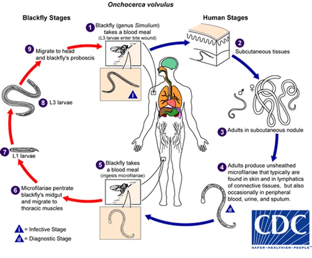
    Onchocerciasis is spread by female blackflies (Simuliumgenus) that breed in swiftly flowing streams (hence, the term “river blindness”). Many blackfly bites are needed before disease develops. Infective larvae inoculated into the skin during the bite of a blackfly develop into adult worms in 12 to 18 months. Adult female worms may live up to 15 years in subcutaneous nodules. Females are 33 to 50 cm long; males are 19 to 42 mm long. Mature female worms produce microfilariae that migrate mainly through the skin and invade the eyes.


    Symptoms and Signs of Onchocerciasis


    Onchocerciasis typically affects

    • Skin (nodules, dermatitis)
    • Eyes


    Nodules

    The subcutaneous (or deeper) nodules (onchocercoma) that contain adult worms may be visible or palpable but are otherwise asymptomatic. They are composed of inflammatory cells and fibrotic tissue in various proportions. Old nodules may caseate or calcify.

    Patients may have enlargement of inguinal, femoral, or other lymph nodes. Localized swelling of the genitalia and inguinal hernias can develop.


    Skin disease

    Onchocercal dermatitis is caused by the microfilarial stage of the parasite. Intense pruritus may be the only symptom in lightly infected people. 


    Skin lesions usually consist of a nondescript maculopapular rash with secondary excoriations, scaling ulcerations and lichenification, and mild to moderate lymphadenopathy. Other skin abnormalities can include premature wrinkling, atrophy, patchy hypopigmentation, and loss of elasticity. In severe cases, patients may develop folds of atrophic skin in the lower abdomen and upper medial thighs (“hanging groin”).

    Onchocercal dermatitis is generalized in most patients, but a localized and sharply delineated form of eczematous dermatitis with hyperkeratosis, scaling, and pigment changes (Sowdah) is common in Yemen and Sudan.
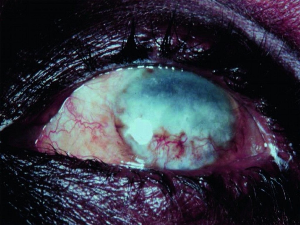
     

    Eye disease

    Ocular involvement ranges from mild visual impairment to complete blindness. Lesions of the anterior portion of the eye include

    • Punctate (snowflake) keratitis (an acute inflammatory infiltrate surrounding dying microfilariae that resolves without causing permanent damage)
    • Sclerosing keratitis (an ingrowth of fibrovascular scar tissue that may cause subluxation of the lens and blindness)
    • Anterior uveitis or iridocyclitis (which may deform the pupil)

    Chorioretinitis, optic neuritis, and optic atrophy may also occur.


    Diagnosis of Onchocerciasis 


    • Microscopic examination of skin snips or biopsies
    • Slit-lamp examination of the cornea and anterior chamber of the eye

    Demonstration of microfilariae in skin snips or biopsies is the traditional diagnostic method for onchocerciasis; multiple samples are usually taken (see table Collecting and Handling Specimens for Microscopic Diagnosis of Parasitic Infections). PCR-based methods to detect parasite DNA in skin samples are more sensitive than standard techniques but are available only in research settings. 


    Microfilariae may also be visible in the cornea and anterior chamber of the eye during slit-lamp examination.


    Antibody detection is of limited value; there is substantial antigenic cross-reactivity among O. volvulus and other filaria and different helminths, and a positive serologic test does not distinguish between past and current infection.

    Palpable nodules (or deep nodules detected by ultrasound or MRI) can be excised and examined for adult worms, but this procedure is rarely necessary.


    Treatment of Onchocerciasis 


    • Ivermectin

    Ivermectin, the primary therapeutic option, reduces microfilariae in the skin and eyes and decreases production of microfilariae for many months. It does not kill adult female worms, but cumulative doses decrease their fertility. Ivermectin is given as a single oral dose of 150 mcg/kg, repeated at 6- to 12-month intervals. The optimal duration of therapy is uncertain. Although treatment could theoretically be continued for the life span of female worms (10 to 14 years), it is usually stopped after several years if pruritis has resolved and no evidence of microfilariae is detected by skin biopsy or eye examination.


    Adverse effects of ivermectin are qualitatively similar to those of diethylcarbamazine (DEC) but are much less common and less severe. DEC is not used for onchocerciasis because it can cause a severe hypersensitivity (Mazzotti) reaction to released filarial antigens, which can further damage skin and eyes and lead to cardiovascular collapse. Mild Mazzotti reactions are a frequent complication during treatment, and severe reactions can be managed with low-dose corticosteroids rapidly tapered (1). 

    Before treatment with ivermectin, patients should be assessed for coinfection with Loa loa, another filarial parasite, if they have been in areas of central Africa where both parasites are transmitted because ivermectin can cause severe reactions in patients with heavy Loa loa coinfection.


    Sources & Credit:

    https://www.msdmanuals.com/professional/infectious-diseases/nematodes-roundworms/onchocerciasis-river-blindness?query=Onchocerciasis%20(River%20Blindness)

    https://www.msdmanuals.com/home/infections/parasitic-infections-roundworms-nematodes/overview-of-filarial-worm-infections

    Rating: 5 out of 5.

    5,138 hits

    Leave a comment

    𝙷𝚘𝚠 𝚖𝚎𝚍𝚒𝚌𝚒𝚗𝚎 𝚊𝚗𝚍 𝚑𝚎𝚊𝚕𝚝𝚑𝚌𝚊𝚛𝚎 𝚊𝚏𝚏𝚎𝚌𝚝 𝚞𝚜 𝚒𝚗 𝚝𝚑𝚎 𝚜𝚖𝚊𝚕𝚕𝚎𝚜𝚝 𝚘𝚏 𝚠𝚊𝚢𝚜 𝚕𝚎𝚊𝚍𝚒𝚗𝚐 𝚝𝚘 𝚋𝚒𝚐𝚐𝚎𝚛 𝚒𝚖𝚙𝚊𝚌𝚝𝚜 𝚊𝚗𝚍 𝚕𝚒𝚏𝚎-𝚌𝚑𝚊𝚗𝚐𝚒𝚗𝚐 𝚌𝚘𝚗𝚜𝚎𝚚𝚞𝚎𝚗𝚌𝚎𝚜! 𝚄𝚕𝚝𝚒𝚖𝚊𝚝𝚎𝚕𝚢, 𝚌𝚑𝚊𝚗𝚐𝚒𝚗𝚐 𝚠𝚑𝚊𝚝 𝚠𝚎 𝚌𝚊𝚕𝚕 ‘𝚑𝚎𝚊𝚕𝚝𝚑𝚌𝚊𝚛𝚎.’

    “Copyright [2024], [2025], [2026], [Scriveners], [Scriveners Online], All Rights Reserved. Any unauthorised duplication or use of this material is strictly prohibited.”

    ‘𝙰 𝚙𝚛𝚘𝚞𝚍 𝚖𝚎𝚍𝚒𝚌𝚊𝚕 𝚜𝚝𝚞𝚍𝚎𝚗𝚝’𝚜 𝚒𝚗𝚒𝚝𝚒𝚊𝚝𝚒𝚟𝚎 𝚊𝚗𝚍 𝚘𝚠𝚗𝚎𝚛𝚜𝚑𝚒𝚙!’‘